

A recent case study report describes the non-surgical repair of a left internal mammary artery (IMA) branch pseudoaneurysm. This complication occurred after a chest surgery and can in some cases lead to death. The study, titled “Pseudoaneurysm of a branch of left internal mammary artery: a late and potentially fatal complication after redo-sternotomy: a case report” appeared in the March 18th issue of the journal Interactive and Cardiovascular Thoracic Surgery.
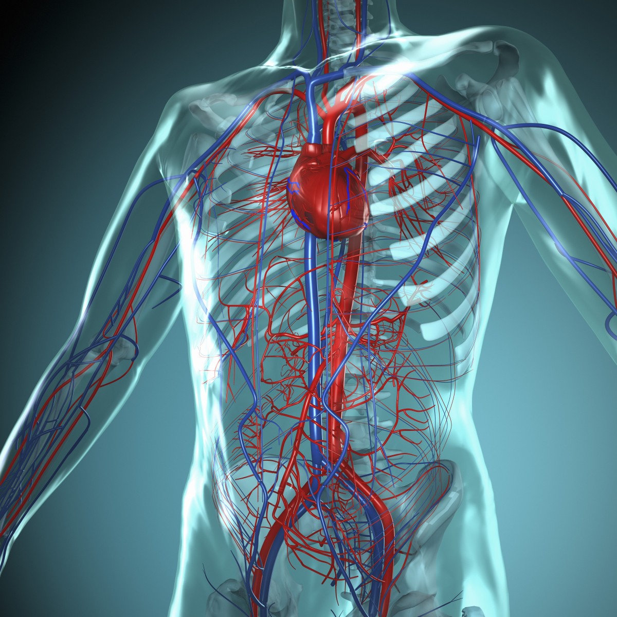
The IMA is also called the internal thoracic artery. It supplies the anterior chest wall and the breasts. A pseudo aneurysm is also called a “false aneurysm.” It is a collection of blood (hematoma) that forms due a leaking hole in an artery.
 The case study report, led by Fabio Falconieri of Royal Brompton and Harefield Hospital NHS Foundation Trust in London, focused on a 78-year old woman with a (IMA) branch pseudo aneurysm that had appeared a few weeks after a redone chest surgery to repair the mitral valve, a flap in the heart. According to the study authors, the pseudo aneurysm that occurred was uncommon, but could have lead to serious complications. The authors stated that “Post-sternotomy pseudo aneurysms of the internal mammary arteries (IMAs) and their branches are rare and often present with rupture-associated haemothorax and haemodynamic instability.” The researchers warned that “Finally, failure to identify and treat these complications may result in rupture, bleeding and death.”
The investigators identified the pseudoaneurysm using computerized axial tomography (CT) scanning, stating “Evaluation with angiography or computed tomography scan with 3D reconstruction can establish the diagnosis and lead the way towards treatment.”
They then used a non-surgical procedure to embolize the pseudoaneurysm, a nonsurgical procedure that selectively blocks a blood vessel. In this case, it was used to stop the bleeding.
The non-surgical treatment was successful in this patient. According to the authors, “The patient was then discharged home and followed up 6 weeks later in our outpatient clinic where she presented in good condition and the parasternal swelling was reduced in calibre.”
Non-surgical treatments such as the one described help facilitate faster recovery and cause less pain to the patient. This case study gives a good example of how a cardiovascular condition can be treated non-surgically. As non-invasive treatments become more and more common, treatments for cardiovascular problems are less damaging and life-impairing for patients.
More studies of this type of intervention are of course needed to determine whether they may be used routinely.
