
A team of investigators at the UT Southwestern Medical Center in Dallas, Texas have found that prolonged use of left ventricular assist devices (LVAD) by heart failure patients may induce heart muscle regeneration of by way of preventing oxidative damage to a cell-regulating mechanism.
LVADs are the mechanical pumps sometimes implanted in patients awaiting heart transplants. The LVAD takes over from the damaged heart in the task of pumping blood for circulation throughout the body.
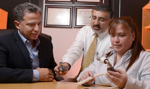
The study, entitled “Human Ventricular Unloading Induces Cardiomyocyte Proliferation“ (J Am Coll Cardiol. 2015;():. doi:10.1016/j.jacc.2014.12.027) published online first in the Journal of The American College of Cardiology, led by senior author UT Southwestern Assistant Professor of Internal Medicine Dr. Hesham Sadek with Diana C. Canseco PhD, Wataru Kimura PhD, Sonia Garg MD, Shibani Mukherjee PhD, Souparno Bhattacharya MS, Salim Abdisalaam PhD, Sandeep Das, MD; Aroumougame Asaithamby, PhD; and Pradeep P.A. Mammen MD, variously of the Department of Internal Medicine, Department of Psychiatry, and Department of Radiation Oncology at UT Southwestern Medical Center, investigated pre- and post-LVAD heart muscle samples in ten heart failure patients, examining paired tissue samples for DNA damage and cell proliferation markers.
The coauthors note that adult mammalian hearts are incapable of meaningful regeneration after substantial cardiomyocyte loss, due primarily to inability of adult cardiomyocytes to divide.
However, the UT Southwestern research team recently showed that mitochondria-mediated oxidative DNA damage is an important regulator of postnatal cardiomyocyte cell cycle arrest, but it is not known whether mechanical load also plays a role in this process. The scientists reasoned that the postnatal physiological increase in mechanical load contributes to the increase in mitochondrial content, with subsequent activation of DNA damage response (DDR) and permanent cell cycle arrest of cardiomyocytes.
The study’s main objective was to test the effect of mechanical unloading on mitochondrial mass, DDR, and cardiomyocyte proliferation. IN their investigation, the researchers examined the effect of human ventricular unloading after implantation of left ventricular assist devices (LVADs) on mitochondrial content, DDR, and cardiomyocyte proliferation in ten matched left ventricular samples collected at the time of LVAD implantation (pre-LVAD) and at the time of explantation (post-LVAD).
They found post-LVAD hearts showing up to a 60 percent decrease in mitochondrial content and up to a 45 percent decrease in cardiomyocyte size compared with pre-LVAD hearts. Moreover, they were able to quantify cardiomyocyte nuclear foci of phosphorylated ataxia telangiectasia mutated protein, which is an upstream regulator of the DDR pathway, observing a significant decrease in numbers of nuclear phosphorylated ataxia telangiectasia mutated foci in the post-LVAD hearts.
Finally, the researchers examined cardiomyocyte mitosis and cytokinesis, finding a statistically significant increase in both phosphorylated histone H3positive, and Aurora Bpositive cardiomyocytes in the post-LVAD hearts. They emphasize that these results were driven by statistical significance in hearts exposed to longer durations of mechanical unloading, concluding that prolonged mechanical unloading induces adult human cardiomyocyte proliferation, possibly through prevention of mitochondria-mediated activation of DDR.
This study builds on earlier work with mouse models demonstrating that newborn mammalian hearts are capable of a strong, regenerative injury response by cell division activation. The earlier studies had demonstrated how injury response ability is lost due to circulation changes after birth that result in a more oxygenated environment in the heart, and ultimately in oxidative damage to cellular machinery controlling heart-muscle regeneration.
The UT Southwestern investigators reasoned that by assisting the damaged heart, LVADs would alleviate oxidative damage occurring within heart-muscle cells.
 “We looked at markers of what is called the DNA damage response in cardiomyocytes (heart-muscle cells) of these patients,” says Dr. Sadek. “The response is composed of a cascade of proteins that is activated in response to DNA damage and in turn shuts off the ability of cardiomyocytes to divide. We found that patients who were on LVAD for more than six months had significantly decreased levels of DNA damage response.”
Examining the paired tissue samples for markers of cell division, the investigators found that patients who had been on LVADs for six months or longer showed a significant increase in cardiomyocyte cell proliferation — indeed nearly triple the amount.
“This result shows that patients with mechanical assist devices have the ability to make their muscle cells divide,” says Dr. Sadek. “And the obvious question now is, ‘Are these hearts regenerating? Could LVADs be used as a cure for heart failure?'”
Some 6 million people in the United States are currently living with heart failure, according to the American Heart Association, with incidence of the condition expected to soar over the next two decades with an aging population ages and improvements in heart-attack treatments leading to more people surviving heart attacks.
 Study co-senior author Dr. Pradeep Mammen, Associate Professor of Internal Medicine at UT Southwestern, comments that “Putting in a mechanical pump rests the heart and apparently sends a signal to make new heart cells. This is the first time that this phenomenon has been shown to occur in human heart failure.”
Dr. Sadek says the next step in the team’s research will be to document that the cell division they observed produces viable heart tissue and a stronger pumping function.
 Dr. Joseph Hill, Professor of Internal Medicine and Molecular Biology and UT Southwestern Chief of Cardiology calls this new research “exciting,” observing that “Dr. Sadek’s findings raise the prospect of reawakening otherwise quiescent cardiac muscle cells, coaxing them into regenerating new and healthy cells. This has been an over-arching objective of the field for many years. The next step will be to leverage these exciting results to rebuild the failing heart.”
 “This is an exciting advance. We have a long way to go, but hopefully this study will be an important first step toward uncovering methods of promoting myocardial recovery,” says Dr. Mark Drazner, Professor of Internal Medicine and Medical Director of the UT Southwestern Heart Failure, LVAD, and Cardiology Transplant Program.
Dr. Sadek and Dr. Mammen are members of UT Southwestern’s newly-established Hamon Center for Regenerative Science and Medicine, which was made possible by the gifting of a $10 million endowment from the Hamon Charitable Foundation. The Center’s goal is to understand the basic mechanisms for tissue and organ formation, and then to use the knowledge to regenerate, repair, and replace tissues damaged by aging and injury.
 Dr. Eric Olson, Professor of Molecular Biology and Chairman of the Department of Molecular Biology, and head of the Hamon Center, notes: “This is a fascinating study and a wonderful example of the translation of a discovery in basic science to an important new insight into human disease. Dr. Sadek’s work provides fresh new ideas for stimulating heart regeneration, which is one of the biggest challenges in cardiovascular medicine today.”
Sources:
UT Southwestern Medical Center
Journal of the American College of Cardiology
Image Credits:
UT Southwestern Medical Center
